Pearls of Cleaning and Sterilizing Surgical Instruments

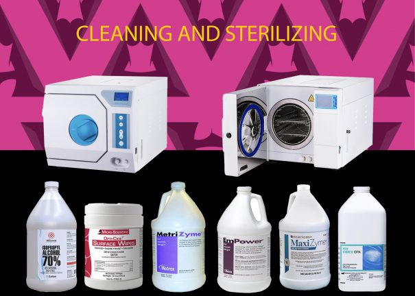
Cleaning is the removal of foreign material (soil, organic material) from objects and is normally accomplished using water with detergents or enzymatic products (MaxiZyme, Metrizyme, Empower).  Thorough cleaning is required before disinfection and sterilization. 

Disinfection describes a process that eliminates pathogenic microorganisms, except bacterial spores, on inanimate objects.

Sterilization describes a process that destroys or eliminates all forms of microbial life. 

Unlike sterilization, disinfection is not sporicidal.

The term germicide includes both antiseptics and disinfectants. Antiseptics are germicides applied to living tissue and skin; disinfectants are antimicrobials applied only to inanimate objects.  In general, antiseptics are used only on the skin and not for surface disinfection, and disinfectants are not used for skin antisepsis because they can injure skin and other tissues.

The effective use of disinfectants is part of a multi-barrier strategy to prevent health-care–associated infections.  Surfaces are considered noncritical items because they contact intact skin.  Use of noncritical items or contact with noncritical surfaces carries little risk of causing an infection in patients or staff.  Nonetheless, cleaning and disinfecting surfaces is common practice and should be routinely practiced.

Surgical instruments should be presoaked to prevent drying of blood, and to soften and remove blood/fat from the instruments.  Cleaning may be done manually, by rubbing/scrubbing the soiled area with a brush to remove any blood, fat, or any other organic material from the surface, using water with detergents or enzymatic products, or mechanically, by using automatic cleaners such as ultrasonic cleaners, washer-de-contaminators, washer-disinfectors, and washer-sterilizers.

For instrument cleaning, a neutral or near-neutral pH detergent solution containing enzymes commonly is used because such solutions are active on blood/fat/carbohydrates providing good soil removal.  Enzymatic cleaners are not disinfectants and as with all chemicals, enzymes must be rinsed from the equipment.  Enzyme solutions should be used in accordance with manufacturer’s instructions, which include proper dilution of the enzymatic detergent and contact with equipment for the amount of time specified on the label.

Many disinfectants (alcohol, glutaraldehyde (Cydex), hydrogen peroxide) are used alone or in combinations. 

Steam sterilization, moist heat in the form of saturated steam under pressure, is the most widely used and the most dependable method of surgical sterilization.  Steam sterilization is nontoxic, inexpensive, rapidly microbicidal, sporicidal, and rapidly heats and penetrates fabrics.  The basic principle of steam sterilization, as accomplished in an autoclave, is to expose each item to direct steam contact at the required temperature and pressure for the specified time. 

There are four (4) parameters of steam sterilization: steam, pressure, temperature, and time.  The ideal steam for sterilization is dry saturated steam and entrained water.  Pressure serves as a means to obtain the high temperatures necessary to quickly kill microorganisms.  Specific temperatures must be obtained to ensure the microbicidal activity.  The two common steam-sterilizing temperatures are 121°C (250°F) and 132°C (270°F).  These temperatures (and other high temperatures) must be maintained for a minimal time to kill microorganisms.  At constant temperatures, sterilization times vary depending on the type of item (metal versus rubber, plastic), whether the item is wrapped or unwrapped, and the sterilizer type.

The two basic types of steam sterilizers (autoclaves) are the gravity displacement autoclave and the high-speed pre-vacuum sterilizer.  Refer to the autoclave’s manual of operation for the type of steam sterilizer and the minimum cycle times for steam sterilization cycles.

Flash steam sterilization refers to sterilization of an unwrapped object and it depends on the type of sterilizer and the type of item (porous vs non-porous items).  Although the wrapped method of sterilization is preferred, correctly performed flash sterilization is an effective process for the sterilization of critical medical devices.

Flash sterilization is a modification of conventional steam sterilization in which the flashed item is placed in an open tray to allow for rapid penetration of steam.  It is not recommended as a routine sterilization method because of the lack of timely biological indicators to monitor performance, absence of protective packaging following sterilization, possibility for contamination of processed items during transportation to the operating rooms, and the sterilization cycle parameters (time, temperature, pressure) are minimal.

Flash sterilization is considered acceptable for processing cleaned patient-care items that cannot be packaged, sterilized, and stored before use.  It also is used when there is insufficient time to sterilize an item by the preferred package method.  Refer to the autoclave’s manual of operation for the type of steam sterilizer and the minimum cycle times for flash steam sterilization cycles.

Taking the time, putting the effort and following the appropriate guidelines of cleaning and sterilization will ensure the safety of all, patients and medical personnel. 

Cleaning and sterilization of surgical instruments and disinfection of surfaces = Patient Safety!  Connect with us at dravellanet.com.


Circles, Xs, Colors: The Surgical Map


Happy Thursday!  It’s almost time to relax, enjoy the weekend and plan your next surgery.  In previous blogs, I have talked about how and where to place incisions, how to take aesthetic photographs and the importance of planning.  Today I start by saying: The planning of a surgery is critical in a liposuction surgery.  Part of the planning is the pre-operative marking of the patient.  I refer to markings as a surgical map.  The areas to be treated should be marked accordingly using a permanent marker (I have tried various brands but Sharpies work best).  I use specific anatomical landmarks to start drawing my map, use concentric circles to enclose the areas I will be working on, and finalize with Xs of different colors to emphasize the larger size of an area compared to its counterpart.  Here is a video that shows you how I draw my surgical map.

Each surgical map belongs to and should de drawn by each cosmetic specialist.  However you plan it and draw it, it’s entirely up to you.  What’s important is the execution of the plan following a specific and well drawn surgical map. 

Connect with us at dravellanet.com.

My Unforgettable Experience at the First Launch of Beautifill in Latinamerica


A few months ago, at the end of 2020, while doing a training in the United States, I received an international call with an invitation to participate in a Beautifill Training in Central America.  Beautifill is a diode laser from Alma Lasers used for liposuction and fat transfer.  I have been a Beautifill trainer in the US for many years training doctors around the country on the operation and advantages on this equipment.  I had already been in conversation with Alma Lasers since the end of 2019 about collaborating with the international market offering Beautifill Trainings.  The delays that occurred due to the pandemic in 2020, forced face-to-face surgical trainings to be postponed to 2021.  Upon receiving this call, I accepted immediately, as my interest was to enter the Spanish-speaking market as a trainer for this laser equipment.  We immediately began planning for the first Beautifill launch in Latin America to be carried out in Costa Rica, with the local distributor, H&B. 

I had the opportunity to participate in three days of face-to-face training with live surgeries, demonstrating Beautifill for the use in laser liposuction and skin tightening and fat transfer.  On the first day, we trained the clinic SKN, owners of the first Beautifill laser in San José, Costa Rica.  We were able to complete three cases of laser liposuction and fat transfer, including liposculpting of the abdomen and flanks and fat transfer to buttocks, breasts and face.  On the second day, we trained specialists, plastic surgeons and dermatologists, and on the third day we trained aesthetic doctors, offering them, on both days, a talk to discuss the equipment, the operation and advantages, as well as the opportunity to participate in live operations.  All specialists, experts in the technique of liposuction and fat transfer, were completely satisfied with the quality of the laser and its ease of operation and the advantage of offering their patients optimal results using state-of-the-art equipment.  

My experience was enriching as I had the opportunity for the first time, in my native Spanish language, to offer face-to-face Beautifill trainings and share my knowledge with Spanish-speaking colleagues.  Additionally, being able to share state-of-the-art technology with highly skilled and specialized colleagues in the field of liposuction and fat transfer surgery resulted in enriching professional discussions for everyone in attendance, including myself.  The kindness and hospitality of the H&B team was excellent.

Here I share a video of this experience and the participants of the first launch of Beautifill in Latin America, San José, Costa Rica.

Pearls of Wisdom: Abdominal, Waist and Back Lipo Incisions

We blogged about Surgical Aesthetic Photography and the importance of measuring and weighing the patient prior to surgery, now we need to address how surgical planning is imperative for the success of any surgery.  The size, placement and number of incisions is a critical initial step in the planning of any aesthetic surgery.  Liposuction and fat transfer procedures include multiple small incisions placed in key areas ensuring precision and optimization of technique and results.  Enclosed some pearls of wisdom when doing liposuction of the abdomen, waist and back.

  • Incisions are the access points of liposuction cannulas into the subcutaneous fat.
  • Incisions allow post-operative drainage of blood-tinged anesthetic solution, minimizing tenderness, swelling, and edema.
  • The size of an incision will depend on the size of the cannula(s) to be used: an incision should be sufficiently large to permit easy access and avoid friction with the skin that could potentially cause post-traumatic hyperpigmentation.
  • Incision placement will depend mainly on accessibility and aesthetics: the need to minimize the visibility of potential scars and maximize results.
  • Accessibility of placement is crucial as the idea is to have an incision placed in a location that has the most access to the areas to be treated.  
  • The aesthetics of placement is mostly for the patient as most patients prefer to have very small, imperceptible, hidden scars.
  • The number of incisions required will depend on the size of the areas to be treated.  
  • Not all patients will have the same number of incisions in the same location; this will vary depending on the size of the patient and the areas to be treated.  Patients will ALWAYS ask about the incisions.
  • The average incisions per area is:  abdomen: 3-6; waist: 1-2; love handles: 1-2; bra-line: 1-2; upper back: 1-2.  If the entire abdomen, waist and back are to be treated, a strategic placement of the incisions will cover multiple areas.
  • The more versatility of access, the more areas than can be treated, and the less incisions needed.
The green “X”s shown in the vectors refer to approximate locations of incision placement.

We all want to have good, acceptable and beautiful results for all our patients.  But before we achieve this goal, we need to PLAN, PLAN, PLAN.  Start by planning the number and placement of incisions!  For more information connect with us at dravellanet.com.

Posts navigation